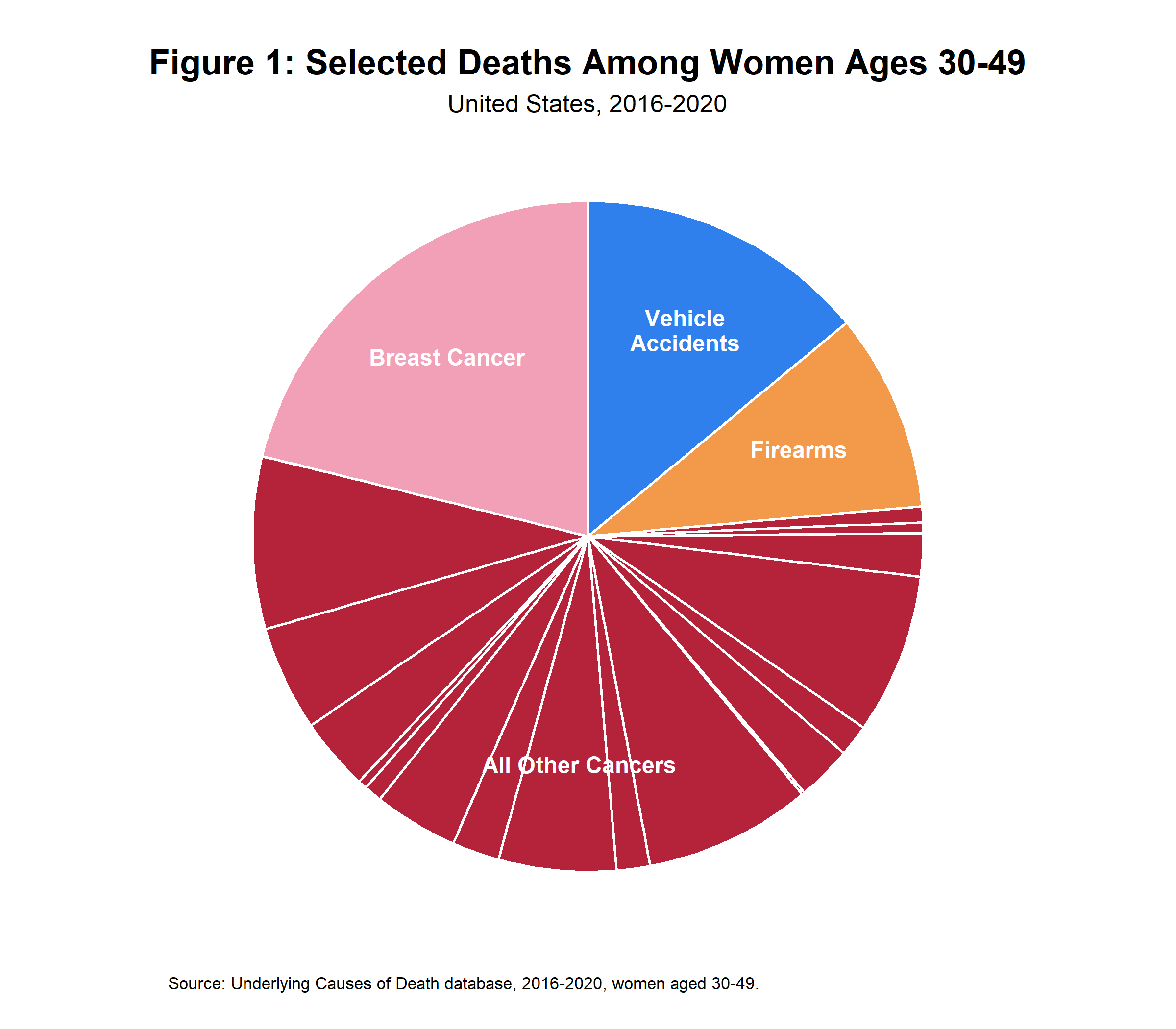
Someone in your life has surely been touched by breast cancer. One in eight women will get it during their lifetime. It kills more women in their thirties and forties than guns, vehicles, or any other form of cancer. As a woman, whatever you fear, you should probably fear this more.
Does it have to be this way? Treatment is certainly advancing, with biomarker-directed therapies and antibody-drug conjugates expanding the option set and slowing disease progression by months.
But for the young or middle-aged woman who hasn’t been affected, there is low-hanging fruit: screening. Screen smarter and screen earlier. Rather than relying on stale guidance which use outdated technology and deploy them too late (“mammograms starting at forty”), take control of your health. Use modern tools like an MRI, and use them in your thirties and even late twenties.
Cancer is horrible, in how it breaks families, drains savings, and debilitates its victims. So, get to it before it gets to you.
What they want you to do
The standard medical advice is that women should get mammograms starting at the age of forty. This has been the recommendation for decades now; the American Cancer Society first recommended it in 1983 after successful trials in the 1960s. Supposedly the mammogram offers a good balance of efficacy, risks, and costs compared to, for instance, the MRI.
I disagree, and I wrote this article to convince you too. As background, this article is inspired by the book on pregnancy Expecting Better, by Emily Oster, an economist. (I too am an economist.) Expecting Better questions and investigates the established medical advice from the ground-up, by looking at the underlying studies and evaluating the evidence they provide. And so this article is me bringing that same ethos to breast cancer.
Let’s return to the mammogram recommendation. I (sort of) understand it. The bodies that issue them, e.g. the US Preventive Services Task Force, have a tough job. They have to balance everyone’s tradeoffs and preferences. Some patients have strong mental fortitude and can tolerate false positives; some cannot. Some patients are wealthy and can afford out-of-pocket screening; some are not. And so on. In having to cater to such a disparate group — especially under a “do no harm” mindset — they issue a recommendation that is extremely conservative.
But not all women are the same. Some can tolerate false positives and extra costs if they come with more detected true positives. Those women can and should go beyond the lowest common denominator advice, and take more active roles in their health.
Those women are the intended readers for my article.
And indeed, it is important to understand that this article leaves the beaten path. There is no single study that compares the recommendations herein to the default ones. Such a study would involve a randomized controlled trial that tracks tens of thousands of patients for decades, which is as slow as it is costly. Instead, this article tries to stitch together several studies where each individually provides a useful nugget of information to the broader question. This may be bold, and it may be foolish. But remember that waiting for that perfect study, and equating absence of evidence with evidence of absence in the meantime, can be just as foolish. For breast cancer does not wait.
MRIncredible
The first and most important thing to understand is that MRIs are superior to mammograms in detecting cancer. In study after study, they pick up more invasive cancers than mammograms; and with similar or even lower false positive burdens.
May the best scan win
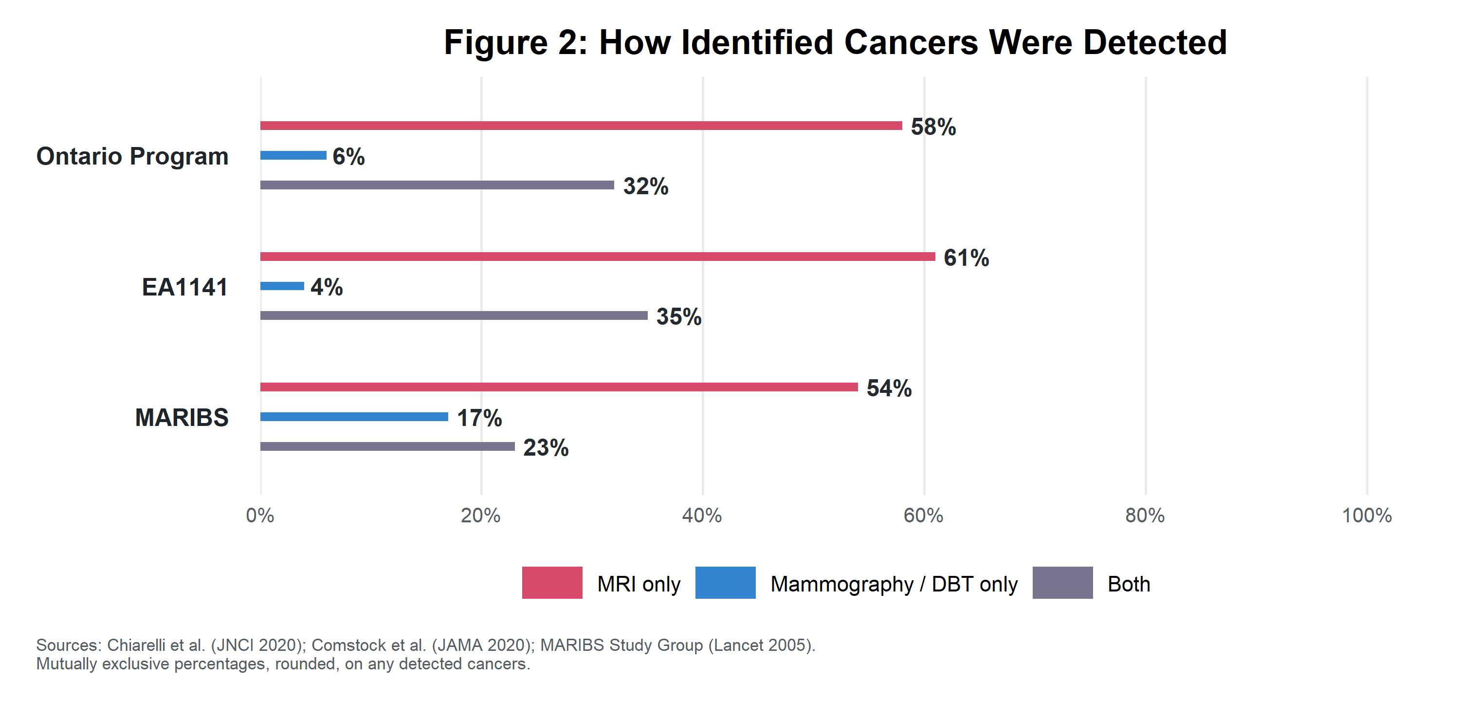
A straightforward way to show that MRIs are more effective than mammograms is to simply compare them head-to-head. In such studies, patients are given both MRIs and mammograms, and detection rates are compared within the same person. This tells us immediately whether one technique is better at catching cancers. And here, a battery of studies show that the MRI catches roughly twice as many cancers as mammography. Here are three such examples:
- An Ontario screening program of 9,000 women finds that MRIs catch 90% of total identified cancers to mammography’s 38%.
- The EA1141 trial, which focuses on the abbreviated MRI specifically, finds the abbreviated MRI catches 96% of the identified cancers to mammography’s 39%.
- The UK MARIBS trial, which includes a focus on women with BRCA1 gene mutations, shows that MRI scores 77% to mammography’s 40%; and a stark 92% to 23% in the BRCA1 subgroup.
Not all cancers
The initial results seem stark, but a common criticism of more sensitive cancer screening routines is that not all tumors matter. Many tumors are benign in perpetuity, and so the technique that finds more of these benign tumors is not actually useful and possibly harmful.
But there is good evidence to show that MRIs find tumors that matter, i.e. ones that become problematic. These come from randomized controlled trials, wherein some patients are assigned to receive MRIs and others to mammograms. If those patients are tracked over time, they can be directly compared for incidence of malignant cancer.
The highest-quality trial is DENSE, which invited 40,000 women in the Netherlands with extremely dense breasts and a negative screening mammogram into a study, and randomized them into two pathways: one based on mammography, and one with a supplemental MRI. Women in the study were then screened, and cancers found during the screening were treated.
Then — and this is the key — the authors simply waited and watched. Over the next two years, would women in the two groups suffer surprise bouts with breast cancer differently? Which group stayed healthier? Formally, their metric is an “interval cancer,” i.e. cancers that emerge in the interval between routine checkups. These are cancers that generally matter, as they are the ones that generate sufficient irregularities to require immediate care.
Women in the MRI arm had half the interval cancer rate of those in the mammogram arm, i.e. 0.25% vs 0.50%. And in fact, if anything, this is understating the results. Not all women in the MRI arm actually accepted the MRI; and they are still included in that 0.25% number. For the women who actually completed the MRI, the interval cancer rate is a tiny 0.08%.
This is the strongest single piece of evidence in support of MRIs: women given them were healthier. Of course, critics will urge caution from drawing lessons. They will say that women with dense breasts constitute a subpopulation where MRIs work better than mammograms (though we’ll return to this point later), or that interval cancers are not always fatal. So yes, sure, your mileage may vary. But paired with the many head-to-head studies, the evidence looks increasingly promising.
False alarm about false alarms
The second major criticism of any robust screening program is the false positive rate. Let’s hold aside the paternalism of such a critique — which I’ll also return to later — and show that, if anything, it bites harder on mammograms than MRIs.
The best data points come from the second major randomized controlled trial, known as the FaMRIsc trial (also from the Netherlands). This invited 1,400 women with high familial breast cancer risk into a study, and randomized them to two pathways: an annual MRI and biennial mammogram, or an annual mammogram. As a randomized controlled trial, this study is naturally insightful on direct outcomes. For instance, the MRI arm found cancer at a much higher rate than the mammogram arm, at 1.4% of patients to 0.5%. Owing to the smaller sample size, it is more limited in long-term outcomes than DENSE, e.g. the study found fewer interval cancers in the MRI arm but not at a statistically significant level. But even so, it did find that the cancers that emerged later in the mammography arm (in incident rounds) were more severe: 63% had spread to lymph nodes, versus only 11% in the MRI arm.
But for our purposes, FaMRIsc is especially insightful on the false positive question. Figure 3 visualizes the two arms and the conditional probability tree of different pathways. In raw numbers, MRIs flag more screens and find more overall false positives compared to mammograms. But per true positive, i.e. defined here per invasive cancer found, MRIs find fewer false positives overall and comparable false positives at the biopsy stage (which is where such false positives are the most costly). These numbers are validated in other and larger trials, e.g. the Ontario study.1
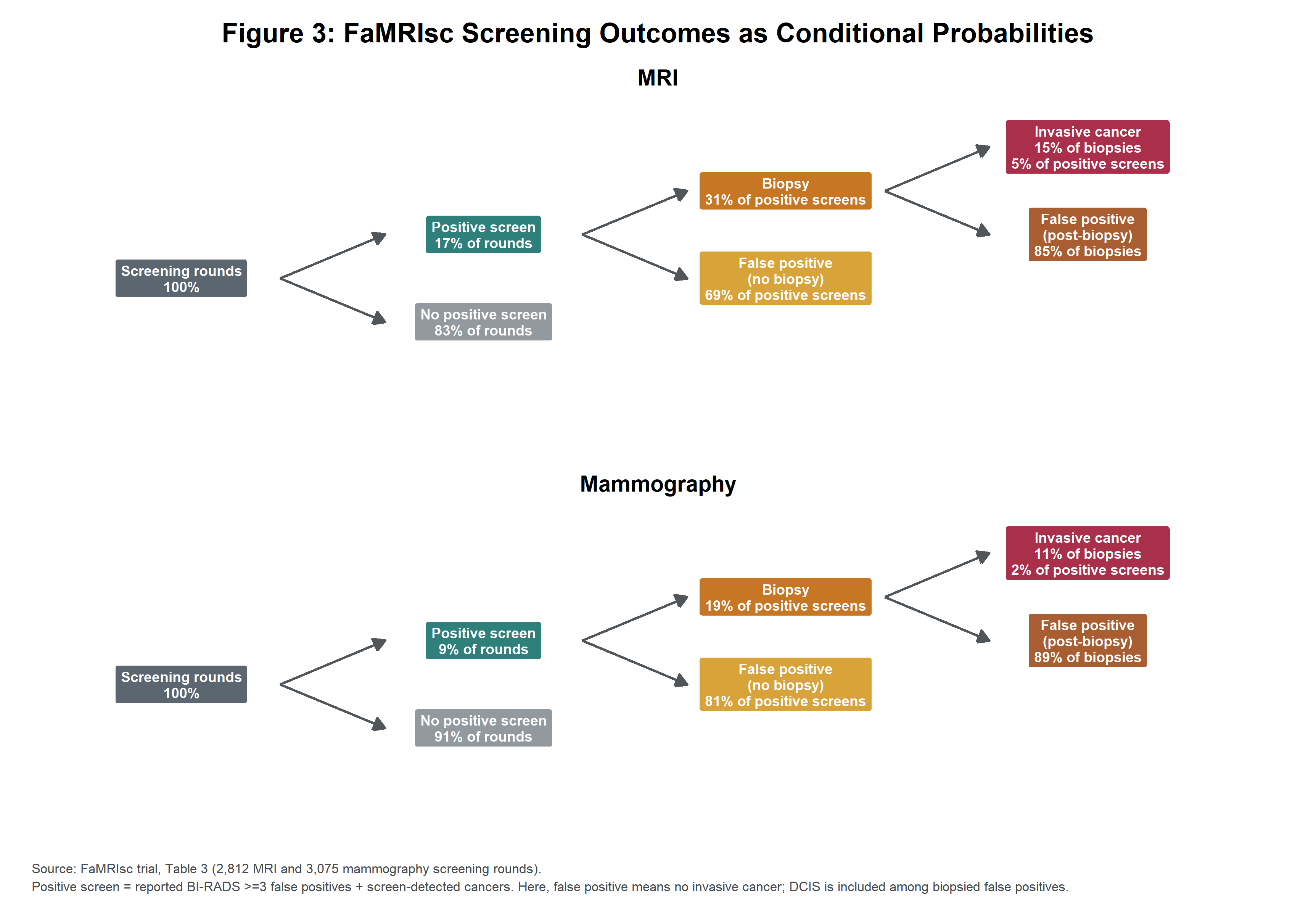
And so if anything, the false positive concern is more apt for mammograms. After all, you should not care about the raw number of false positives; you should care about their prevalence relative to true positives. If you are comfortable with the tradeoff posed by the mammogram, you should be even more comfortable with the one posed by the MRI.
This rounds out the argument for the MRI. It is that rare technology that both catches more concerns, and at a comparable or even higher signal-to-noise ratio, than the incumbent.
Early and often
Picking the right tool is only half the job; the other half is knowing when to use it. Here, I believe the incremental value-add of an elective MRI is highest at young ages (e.g. late twenties and thirties).
This may seem aggressive, especially since many of the studies above are on older women. But breast cancer is not just an old woman’s disease. Indeed, the types of breast cancer that are scariest — such as the triple-negative subtypes (which have more limited treatment options) and HER2-positive subtypes (which are fast-growing) — are over-represented in women under 40. BRCA1 tumors, which are typically high-grade and fast-moving, also cluster in young women.
More specifically, there are four reasons to start at a younger age.
- Once women hit their forties, breast cancer screening becomes routine. While the default tool here is mammography, this at least offers some defense against breast cancer. By contrast, at younger ages, the choice is often between an elective screen and nothing. (If women are offered anything at these younger ages, it is just a physical exam, which the American Cancer Society flatly sees no value in for women of any risk profile and any age.)
- Young women have denser breasts, with markedly more glandular and connective tissue during their reproductive years. By one estimate, roughly 75% of women in their forties have breasts composed largely of denser fibroglandular tissues rather than fatty tissues. This obscures abnormalities on mammograms, but MRIs (for technical reasons, discussed in the Appendix) are much less affected by density. Indeed, the Ontario study breaks out numbers for this subpopulation, and shows that MRIs for younger women had 93% sensitivity to the mammogram’s 43%.
- Early cancers, while rare, are unusually aggressive. This motivates starting any screening regime early, both to lower the mortality risk and to minimize the pain of treatment. Survival rates are unsurprisingly related to the stage in which a cancer is detected: cancer caught when localized has a 99% survival rate; cancer caught when metastatic has a grim 33% survival rate. Moreover, cancer treatment is naturally more invasive and intensive the later the cancer is caught. Here, a matched analysis called MRISC shows that women who detected cancers via MRI-based screening routines were less likely to need chemotherapy than matched controls (39% versus 77%). Although matched analyses are not strong forms of evidence on their own, this finding is consistent with the other studies.
- Once a baseline is established early, subsequent MRIs can more easily identify changes.2 The malignancy of a tumor is related to the speed of its growth, and this can best be identified through repeated measurements over time.
Must be funny, in a rich woman’s world
Life has no free lunches, and indeed MRIs are expensive and time-consuming (not unduly so, but around one or two thousand dollars for 30-40 minutes of scan time). Moreover, they are often not covered by insurance if ordered as an elective procedure.
But don’t let that stop you, for there is another option: a technology called the “abbreviated MRI” (i.e. AB-MRI). An AB-MRI only captures a handful of images, to answer the simple question of whether there is anything worth further investigation. They are much cheaper and quicker, i.e. a few hundred dollars and only a few minutes of scan time.
The Appendix covers the technical details of the procedure; but the most important thing is that they work comparably well to the MRI. The benchmark here is the EA1141 study, which tested AB-MRI directly against the modern standard of mammography (specifically known as digital breast tomosynthesis). In the study, some 1,400 women were given both, and independent radiologists read the results of each.
The results strongly mirrored the comparison between the full MRI and mammogram. The abbreviated MRI detected invasive cancers in 1.2% of the subjects to mammogram’s 0.5%, which is very similar to the full MRI’s detection rate in DENSE and FaMRIsc. The two MRI variants also had similar false positive rates. These numbers are again echoed by other trials, such as the UK BRAID trial, which gave AB-MRIs to women with negative mammograms, and found invasive cancer in 1.5% of them.
The main downside is that AB-MRIs give limited visibility, especially for a physician who wants more evidence before recommending a biopsy. But if something suspicious is found, a woman can always return for the full MRI — one with the kinetic curves, additional images, a full morphologic evaluation, and so on — and only then perhaps a biopsy. Indeed, this mirrors how mammograms are deployed: women are initially offered a quick screening mammogram, and then the full diagnostic mammogram only when something suspicious is detected.
From theory to practice
How does one actually operationalize this advice?
Breast MRIs are not available in the US the way that full body scans are, in an integrated concierge-like facility. (Also remember that most full body scans specifically exclude a breast MRI.) Instead, you typically have to do two independent steps:
- Get a physician to write an imaging order.
- Go to a facility that offers a breast MRI or AB-MRI.
There are three ways to get a physician’s order. For the woman with high breast cancer risk, e.g. 20% lifetime risk, prior chest radiation, qualifying mutations, or unusual family history, your physician will often route you to a high-risk breast clinic; and insurance will often cover these procedures. For the lower-risk woman, you have two choices. You can still advocate for your concerns around breast cancer to your usual physician, e.g. an OB-GYN or family medicine physician, regardless of whether your physician assesses it as medically necessary or elective. Alternatively, a telehealth service will typically issue the order with much less resistance and a nominal fee. At this point, you may want to consider calling insurance to see under which conditions they will cover the procedure, which is typically CPT code 77049.
With this order, the second step is to find a clinic that offers the breast MRI or AB-MRI, and critically one that does not require additional steps like a screening mammogram or an extra questionnaire on patient risk, unless you are willing to do those steps. Every major hospital has an MRI machine, of course; but the out-of-pocket costs can be both high and opaque. (You can still request a “Good Faith Estimate” from these hospitals.) By contrast, clinics are much more reasonable and transparent on costs. This article does not want to promote specific providers, but there are hundreds of such clinics across the US including a few in every major city. Just Google it, or have your AI agent Google it.
The MRI will almost certainly include a reading from a radiologist, who will give a BI-RADS score of 0-6. Scores of 3 (”probably benign”) may warrant follow-up and scores of 4-5 (“suspicious” and “highly suspicious”) definitely do, with support from your primary physician. At this point, such follow-up procedures such as additional imaging and biopsies are much more likely to be classified as medically necessary by insurance.
Finally, you also always have the choice to do nothing, especially with scores of 2-3. The simple knowledge that you have borderline readings can be useful down the road, should it one day be corroborated by another suspicious sign.
Keep calm and carry on
There is much to admire about the medical establishment’s disciplined and deliberate process. But this unintentionally drives its two biggest flaws: conservatism and paternalism. Formal recommendations are issued far too long after the evidence supports them. The bar is set impossibly high for perfection over probability. Those recommendations, in turn, must cater to the lowest common denominator of risk tolerance or mental capacity.
This is most apparent in the concern for false positives, where too often the medical establishment deliberately counsels ignorance to limit false positives. As one such example, in 2009, the US Preventive Services Task Force (the standard setter) changed its flagship recommendation for mammograms to start at forty to the age of fifty; and only restored the age of forty in 2024.3 That is, in recent memory, society made an active choice to receive less information. Indeed, in its discussion of the overdiagnosis problem, it estimates (using admittedly generous estimates): “for every woman who avoids a death from breast cancer through screening, 2 to 3 women will be treated unnecessarily.” And then it makes the decision for you that this tradeoff is unacceptable.
You should not accept this. As a society, we should maximize information and then make good decisions on that information, accounting for the false positive risk. And as an individual, you should get the screening that you want — whether that be an MRI as I believe, a mammogram as the medical establishment recommends, or neither.
So take an active role in your own health. Learn more than the establishment would have you learn. For when it comes to breast cancer, ignorance is not bliss.
Disclaimer
This essay is an argument for taking breast cancer screening more seriously than the current default practice, but you should not consider it to be individualized medical advice. In addition, if you have a breast lump, skin change, persistent pain, or any other acute symptom, seek medical care promptly; this guide is written for asymptomatic individuals. Finally, I have made a good-faith effort to summarize the evidence accurately as of August 2026, but medical evidence and guidelines evolve.
Endnotes
- The Ontario screening program, which is the largest real-world MRI evaluation (with six times as many patients as FaMRIsc), had rates similar to FaMRIsc. The MRI flagged 16% of screening rounds and biopsied 42% of flagged rounds (where FaMRIsc was 17% and 31%), while the mammogram flagged 10% and biopsied 21% of those (where FaMRIsc was 9% and 19%). For both FaMRIsc and the Ontario program and for both the MRI and mammogram arm, approximately one in four biopsies found any cancer and one in seven biopsies found invasive cancer.
- Repeated contrast-enhanced MRIs do leave trace amounts of gadolinium in the body, but this has not been shown to cause any harm to people with normal kidney function. For instance, a study of 1,000 repeatedly imaged patients found no link between exposure and impairments to physical or cognitive function.
- As other examples, the American College of Physicians continues to use fifty as their recommended threshold for mammograms with an option to start at forty; and the American Cancer Society recommends forty-five with an option to start at forty.
Appendix: How an MRI and AB-MRI work
An MRI, and more precisely a contrast-based MRI, works by using a powerful magnet to get tissues to differentially emit energy; and it then images those emissions. More specifically, it leverages two key insights. First, tissues respond more strongly to the magnet when in the presence of gadolinium. Second, malignant tumors have leakier blood vessels. Thus, in an MRI, a gadolinium-based contrast agent is injected intravenously, leaks more strongly into tumor-afflicted tissues than healthy tissues, and subsequently "lights up" more strongly on an image when exposed to the magnet.
Mammography, by contrast, uses low-dose x-ray beams through the breast. Dense tissue attenuates the radiation and appears white, while fatty tissue attenuates less and appears darker; and cancerous tissues are dense. This is why mammograms perform poorest on dense breasts, as tumorous and healthy-but-dense tissues both appear white.
These basic technologies are then wrapped into more elaborate procedures. For instance, virtually all MRIs take images prior to and following the contrast to isolate the differences. Advanced MRIs and modern mammograms (more precisely digital breast tomosynthesis) often take multiple images from multiple angles, to construct a more holistic image of abnormalities. Note that there are other technologies to detect cancer, such as diffusion-weighted imaging — which detects restricted movement of water (common in densely-packed cancerous cells) — but they are deployed far less widely.
The abbreviated MRI is exactly as advertised. Kuhl (2024) notes that there are two modalities: one that focuses on “T1” relaxations (i.e. how quickly nuclei give up energy) and one that focuses on “T2” relaxations (i.e. how quickly nuclei lose coherence from one another). These are typically bundled into tightly-sequenced pre-contrast and post-contrast images, known as the FAST or (sometimes) Ultrafast methodologies. In short, the AB-MRI is just the core of the MRI, without the additional bells and whistles of multiple measurements from different angles, late-stage kinetic curves, diffusion-weighted imaging, and so on. In turn, they are extremely quick, with total scan times of a few minutes, as compared to the typical MRI which can be 30-40 minutes of scan time.
Unlike mammograms, which use radiation, the negative health effects of MRIs and AB-MRIs are muted and short-lived. Most patients suffer nothing — and indeed the DENSE trial reports only 0.1% of its patients suffered adverse effects from MRI screening — and those who do mostly report temporary claustrophobia or a short allergic reaction to gadolinium. There are a few rare cases where contrast-based MRIs specifically can cause issues, e.g. for patients with kidney dysfunction and possibly for pregnant patients, but this holistically makes them better tools for repeated screenings.
References
Core MRI Screening Evidence
- Bakker MF, de Lange SV, Pijnappel RM, et al. "Supplemental MRI Screening for Women with Extremely Dense Breast Tissue." New England Journal of Medicine. 2019;381:2091-2102. https://doi.org/10.1056/NEJMoa1903986
- Veenhuizen SGA, de Lange SV, Bakker MF, et al. "Supplemental Breast MRI for Women with Extremely Dense Breasts: Results of the Second Screening Round of the DENSE Trial." Radiology. 2021;299:278-286. https://doi.org/10.1148/radiol.2021203633
- Saadatmand S, Geuzinge HA, Rutgers EJT, et al. "MRI versus Mammography for Breast Cancer Screening in Women with Familial Risk (FaMRIsc)." Lancet Oncology. 2019;20:1136-1147. https://doi.org/10.1016/S1470-2045(19)30275-X
- Chiarelli AM, Blackmore KM, Muradali D, et al. "Performance Measures of Magnetic Resonance Imaging Plus Mammography in the High Risk Ontario Breast Screening Program." Journal of the National Cancer Institute. 2020;112:136-144. https://doi.org/10.1093/jnci/djz079
- MARIBS Study Group. "Screening with Magnetic Resonance Imaging and Mammography of a UK Population at High Familial Risk of Breast Cancer." Lancet. 2005;365:1769-1778. https://doi.org/10.1016/S0140-6736(05)66481-1
- Kriege M, Brekelmans CTM, Obdeijn IM, et al. "Factors Affecting Sensitivity and Specificity of Screening Mammography and MRI in Women with an Inherited Risk for Breast Cancer." Breast Cancer Research and Treatment. 2006;100:109-119. https://doi.org/10.1007/s10549-006-9230-z
- Saadatmand S, Obdeijn IM, Rutgers EJT, et al. "Survival Benefit in Women with BRCA1 Mutation or Familial Risk in the MRI Screening Study (MRISC)." International Journal of Cancer. 2015;137(7):1729-1738. https://doi.org/10.1002/ijc.29534
Abbreviated MRI and Newer Imaging Evidence
- Comstock CE, Gatsonis C, Newstead GM, et al. "Comparison of Abbreviated Breast MRI vs Digital Breast Tomosynthesis for Breast Cancer Detection Among Women with Dense Breasts Undergoing Screening." JAMA. 2020;323:746-756. https://doi.org/10.1001/jama.2020.0572
- ECOG-ACRIN Cancer Research Group. EA1141 Clinical Trial Results Summary: Comparison of 3D Mammography to Abbreviated Breast MRI. Updated January 2025. Supporting summary, not a peer-reviewed full paper.
- Kuhl CK. "Abbreviated Breast MRI: State of the Art." Radiology. 2024;310:e221822. https://doi.org/10.1148/radiol.221822
- Gilbert FJ, Payne NR, Allajbeu I, et al. "Comparison of Supplemental Breast Cancer Imaging Techniques: Interim Results from the BRAID Randomised Controlled Trial." Lancet. 2025;405:1935-1944. https://doi.org/10.1016/S0140-6736(25)00582-3
Guidelines, Mortality, and Background Data
- US Preventive Services Task Force; Nicholson WK, Silverstein M, Wong JB, et al. "Screening for Breast Cancer: US Preventive Services Task Force Recommendation Statement." JAMA. 2024;331(22):1918-1930. https://doi.org/10.1001/jama.2024.5534
- Siu AL; US Preventive Services Task Force. "Screening for Breast Cancer: US Preventive Services Task Force Recommendation Statement." Annals of Internal Medicine. 2016;164(4):279-296. https://doi.org/10.7326/M15-2886
- Qaseem A, Harrod CS, Balk EM, Etxeandia-Ikobaltzeta I, Crandall CJ; Clinical Guidelines Committee of the American College of Physicians. "Screening for Breast Cancer in Asymptomatic, Average-Risk Adult Females: A Guidance Statement From the American College of Physicians (Version 2)." Annals of Internal Medicine. 2026;179(6):842-856. https://doi.org/10.7326/ANNALS-25-05116
- Oeffinger KC, Fontham ETH, Etzioni R, et al.; American Cancer Society. "Breast Cancer Screening for Women at Average Risk: 2015 Guideline Update From the American Cancer Society." JAMA. 2015;314(15):1599-1614. https://doi.org/10.1001/jama.2015.12783
- American Cancer Society. “American Cancer Society Recommendations for the Early Detection of Breast Cancer.” Last revised December 19, 2023.
https://www.cancer.org/cancer/types/breast-cancer/screening-tests-and-early-detection/american-cancer-society-recommendations-for-the-early-detection-of-breast-cancer.html
- Centers for Disease Control and Prevention, National Center for Health Statistics. Underlying Cause of Death, 2016-2020, Single Race Results. CDC WONDER Online Database. https://wonder.cdc.gov/ucd-icd10-expanded.html
- American Cancer Society. "Survival Rates for Breast Cancer." Revised January 13, 2026. https://www.cancer.org/cancer/types/breast-cancer/understanding-a-breast-cancer-diagnosis/breast-cancer-survival-rates.html
- Checka CM, Chun JE, Schnabel FR, Lee J, Toth H. "The Relationship of Mammographic Density and Age: Implications for Breast Cancer Screening." AJR American Journal of Roentgenology. 2012;198(3):W292-W295. https://doi.org/10.2214/AJR.10.6049
- Nakamura K, McGinley MP, Jones SE, et al. “Gadolinium-based contrast agent exposures and physical and cognitive disability in multiple sclerosis.” Journal of Neuroimaging. 2023;33(1):85–93. doi:10.1111/jon.13057 (https://doi.org/10.1111/jon.13057).
General MRI Context
- Morrow M, Waters J, Morris E. "MRI for Breast Cancer Screening, Diagnosis, and Treatment." Lancet. 2011;378(9805):1804-1811. https://doi.org/10.1016/S0140-6736(11)61350-0